What does the electromagnetic spectrum have to do with the eye?
Lite
Lite is composed of photons that make upwardly electromagnetic waves, which are characterized by wavelength, frequency, and amplitude.
Learning Objectives
Depict the characteristics of lite
Cardinal Takeaways
Key Points
- Calorie-free is equanimous of electromagnetic waves that can travel without a medium, dissimilar sound.
- The behavior of light tin be seen in the behavior of waves and photons, the basic unit of light.
- A wavelength (which varies inversely with frequency ) manifests itself as colour, while wave amplitude is perceived as luminous intensity or effulgence; it is measured by the standard unit of a candela.
- Humans tin can meet calorie-free that ranges between 380 nm and 740 nm, but cannot meet light that is below the frequency of visible red light or above the frequency of visible violet light.
- Light at the crimson end of the visible spectrum has long wavelengths (and is lower frequency), while light at the violet finish has short wavelengths (and is higher frequency).
- Light waves enter the center equally long (cerise), medium (green), and short (blue) waves; the color of an object is the color the object reflects.
Cardinal Terms
- photon: the quantum of light and other electromagnetic energy, regarded as a discrete particle having zero rest mass, no electric charge, and an indefinitely long lifetime
- nanometer: one billionth of a meter; used to express wavelength of calorie-free
- electromagnetic spectrum: the unabridged range of wavelengths of all known radiation consisting of oscillating electric and magnetic fields, including gamma rays, visible light, infrared, radio waves, and X-rays
- wavelength: the length of a unmarried cycle of a wave, equally measured by the distance betwixt one peak or trough of a wave and the next; it corresponds to the velocity of the wave divided by its frequency
Light
Every bit with auditory stimuli, light travels in waves. While the compression waves that compose sound must travel in a medium (consisting of a gas, a liquid, or a solid), light is equanimous of electromagnetic waves and needs no medium. Light can, in fact, travel in a vacuum. The behavior of light can be described in terms of the beliefs of waves and the behavior of the fundamental unit of measurement of calorie-free, the photon: a packet of electromagnetic radiation. A glance at the electromagnetic spectrum shows that visible light for humans is just a small slice of the unabridged spectrum, which includes radiations that nosotros cannot come across every bit calorie-free considering it is below the frequency of visible red light and above the frequency of visible violet light.
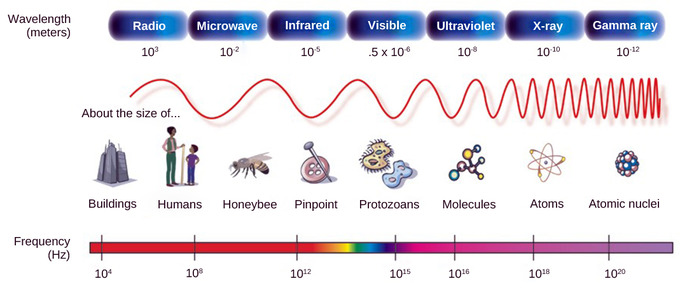
Electromagnetic spectrum: A glance at the electromagnetic spectrum shows that visible light for humans is only a small slice of the unabridged spectrum.
Certain variables are important when discussing perception of low-cal. A wavelength (which varies inversely with frequency) manifests itself equally colour. Light at the red terminate of the visible spectrum has longer wavelengths (and is lower frequency), while light at the violet stop has shorter wavelengths (and is higher frequency). The wavelength of light is expressed in nanometers (nm); i nanometer is one billionth of a meter. Humans perceive light that ranges between approximately 380 nm and 740 nm. Still, some other animals can detect wavelengths outside of the human range. For example, bees see nearly-ultraviolet light in order to locate nectar guides on flowers. Some non-avian reptiles sense infrared light (such as heat that prey gives off).
Moving ridge aamplitude is perceived as luminous intensity or brightness. The standard unit of intensity of lite is the candela, which is approximately the luminous intensity of one common candle.
Light waves travel 299,792 km per second in a vacuum and somewhat slower in various media such as air and water. Those waves arrive at the eye as long (red), medium (green), and short (bluish) waves. The term "white light" is calorie-free that is perceived as white by the human being middle. This upshot is produced by light that stimulates the color receptors in the human being eye equally. The credible colour of an object is actually the color (or colors) the object reflects. Thus a red object reflects the ruby wavelengths in mixed (white) low-cal and absorbs all other wavelengths of light.
Anatomy of the Eye
Many structures in the human centre, such as the cornea and fovea, procedure light and then it tin can exist deciphered by rods and cones in the retina.
Learning Objectives
Explain how eyes have evolved to benefit organisms
Key Takeaways
Key Points
- The cornea and the lens bend low-cal to focus the image on the retina; the iris and pupil regulate the amount of lite inbound the center.
- The aqueous sense of humour maintains the convex shape of the cornea; the vitreous sense of humour supports the lens and maintains the shape of the unabridged eye.
- Presbyopia occurs because the paradigm focuses behind the retina; it is similar to hyperopia (farsightedness), which is caused by an eyeball that is likewise short.
- Myopia (nearsightedness) occurs when an eyeball is elongated; images in the altitude appear blurry, but images nearby are articulate.
- Rods are used for peripheral and nighttime vision; cones are used for daytime and colour vision.
- The fovea is responsible for astute vision because it has a high density of cones.
Key Terms
- rod: a rod-shaped cell located in the outer retina of the eye that is extremely sensitive to calorie-free
- retina: the thin layer of cells at the dorsum of the eyeball where light is converted into neural signals sent to the brain
- cone: jail cell located near the center of the retina that is weakly photosensitive and is responsible for colour vision in relatively vivid light
Anatomy of the Middle
The retina, a sparse layer of cells located on the inner surface of the back of the middle, consists of photoreceptive cells, which are responsible for the transduction of light into nervous impulses. However, calorie-free does not enter the retina unaltered; it must outset pass through other layers that process it so that it can be interpreted past the retina.

Retina: (a) The human eye is shown in cross department. The human eye contains structures, such as the cornea, iris, lens, and fovea, that process light and so information technology can be deciphered by the retina. Other structures like the aqueous humour and the vitreous sense of humor assistance maintain the shape of the eye. (b) A blowup shows the layers of the retina. The retina contains photoreceptive cells. In the retina, light is converted into neural signals sent to the brain.
The cornea, the front transparent layer of the eye, along with the crystalline lens, refract (curve) light to focus the image on the retina. Afterward passing through the cornea, light passes through the aqueous sense of humor, which connects the cornea to the lens. This clear gelatinous mass also provides the corneal epithelium with nutrients and helps maintain the convex shape of the cornea. The iris, which is visible as the colored function of the eye, is a circular muscular band lying betwixt the lens and the aqueous humor that regulates the corporeality of light entering the middle. Lite passes through the eye of the iris, the student, which actively adjusts its size to maintain a abiding level of low-cal inbound the eye. In conditions of high ambient light, the iris contracts, reducing the size of the pupil. In weather condition of low light, the iris relaxes and the student enlarges.
The main function of the lens is to focus lite on the retina and fovea centralis. The lens is a transparent, convex structure located backside the cornea. On the other side of the lens is the vitreous humour, which lets low-cal through without refraction, maintains the shape of the eye, and suspends the fragile lens. The lens focuses and re-focuses light as the eye rests on near and far objects in the visual field. The lens is operated past muscles that stretch it flat or allow it to thicken, changing the focal length of light coming through to focus information technology sharply on the retina. With age comes the loss of the flexibility of the lens; a form of farsightedness chosen presbyopia results. Presbyopia occurs because the image focuses behind the retina. It is a deficit similar to a different type of farsightedness, hyperopia, caused by an eyeball that is likewise short. For both defects, images in the distance are clear, simply images nearby are blurry. Myopia (nearsightedness) occurs when an eyeball is elongated and the image focus falls in front end of the retina. In this example, images in the distance are blurry, but images nearby are clear.
There are two types of photoreceptors in the retina: rods and cones. Both are named for their general advent. Rods, strongly photosensitive, are located in the outer edges of the retina. They detect dim light and are used primarily for peripheral and night vision. Cones, weakly photosensitive, are located almost the heart of the retina. They respond to vivid light; their primary role is in daytime, color vision.

Rods and cones: Rods and cones are photoreceptors in the retina. Rods answer in low lite and can detect merely shades of grayness. Cones reply in intense calorie-free and are responsible for colour vision.
The fovea is the region in the center back of the eye that is responsible for astute (key) vision. The fovea has a loftier density of cones. When you bring your gaze to an object to examine it intently in bright light, the optics orient and so that the object's epitome falls on the fovea. However, when looking at a star in the dark sky or other object in dim light, the object can exist better viewed by the peripheral vision because it is the rods at the edges of the retina, rather than the cones at the middle, that operate better in depression light. In humans, cones far outnumber rods in the fovea.
Transduction of Light
Light is tranduced in rods and cones; visual information is processed in the retina earlier entering the encephalon.
Learning Objectives
Explain retinal processing and the process of transduction of light
Key Takeaways
Central Points
- When lite hits the photoreceptor, the retinal changes shape, which activates the photopigment rhodoposin.
- Primates have total color vision because of the three- cone (trichromatic) organisation; color is a result of the ratio of activity of the three types of cones.
- There are three types of cones with different photopsins: S cones respond to brusk waves; M cones reply to medium waves; L cones reply to lite to long waves.
- If light is not nowadays, neurons are inhibited past rods and cones; once lite is introduced, rods and cones are hyperpolarized, which activates the neurons.
- Activated neurons stimulate ganglion cells, which ship activeness potentials via the optic nerve.
- Horizontal cells can create lateral inhibition, which enhances low-cal and dark contrast in images.
Key Terms
- tonic activity: when photoreceptors get slightly active fifty-fifty when non stimulated by light
- rhodopsin: a light-sensitive pigment in the rod cells of the retina; it consists of an opsin protein spring to the carotenoid retinal
Transduction of Light
The rods and cones are the site of transduction of calorie-free into a neural bespeak. Both rods and cones incorporate photopigments, which are pigments that undergo a chemical modify when they absorb lite. In vertebrates, the principal photopigment, rhodopsin, has ii primary parts: an opsin, which is a membrane protein (in the grade of a cluster of α-helices that span the membrane); and retinal, a molecule that absorbs calorie-free. When light hits a photoreceptor, it causes a shape change in the retinal, altering its structure from a aptitude (cis) form of the molecule to its linear (trans) isomer. This isomerization of retinal activates the rhodopsin, starting a cascade of events that ends with the closing of Na+ channels in the membrane of the photoreceptor. Thus, dissimilar nigh other sensory neurons (which become depolarized by exposure to a stimulus), visual receptors become hyperpolarized and are driven away from the threshold.

Hyperpolarized visual receptors: When lite strikes rhodopsin, the Grand-protein transducin is activated, which in turn activates phosphodiesterase. Phosphodiesterase converts cGMP to GMP, thereby closing sodium channels. As a result, the membrane becomes hyperpolarized. The hyperpolarized membrane does not release glutamate to the bipolar prison cell.

Rhodopsin: (a) Rhodopsin, the photoreceptor in vertebrates, has two parts: the trans-membrane protein opsin and retinal. When light strikes the retinal, it changes shape from (b) a cis to a trans form. The bespeak is passed to a K-poly peptide chosen transducin, triggering a series of downstream events.
Trichromatic Coding
There are three types of cones (with different photopsins) that differ in the wavelength to which they are most responsive. Some cones are maximally responsive to brusk low-cal waves of 420 nm; they are called S cones ("S" for "brusk"). Other cones (M cones, for "medium") answer maximally to waves of 530 nm. A tertiary group (L cones, or "long" cones) responds maximally to light of longer wavelengths at 560 nm. With only 1 blazon of cone, color vision would not be possible; a two-cone (dichromatic) system has limitations. Primates utilise a three-cone (trichromatic) system, resulting in full colour vision.

Rod and cone cells: Human rod cells and the unlike types of cone cells each have an optimal wavelength. However, at that place is considerable overlap in the wavelengths of lite detected.
The color we perceive is a event of the ratio of activity of our three types of cones. The colors of the visual spectrum, running from long-wavelength low-cal to curt are:
- cherry (700 nm)
- orange (600 nm)
- yellow (565 nm)
- green (497 nm)
- blue (470 nm)
- indigo (450 nm)
- violet (425 nm).
Humans accept very sensitive perception of color and can distinguish about 500 levels of effulgence, 200 unlike hues, and 20 steps of saturation; in all, about 2 million distinct colors.
Retinal Processing
Visual signals leave the cones and rods, travel to the bipolar cells, and and then to ganglion cells. A big degree of processing of visual information occurs in the retina itself, before visual data is sent to the brain.
Photoreceptors in the retina continuously undergo tonic activity. That is, they are ever slightly active even when not stimulated by calorie-free. In neurons that exhibit tonic activity, the absence of stimuli maintains a firing charge per unit at an equilibrium; while some stimuli increment firing charge per unit from the baseline, other stimuli decrease firing rate. In the absence of low-cal, the bipolar neurons that connect rods and cones to ganglion cells are continuously and actively inhibited by the rods and cones. Exposure of the retina to low-cal hyperpolarizes the rods and cones, removing the inhibition of their bipolar cells. The now-active bipolar cells in turn stimulate the ganglion cells, which send activeness potentials forth their axons (which leave the eye every bit the optic nerve). Thus, the visual arrangement relies on changein retinal activeness, rather than the absence or presence of activity, to encode visual signals for the brain. Sometimes horizontal cells behave signals from one rod or cone to other photoreceptors and to several bipolar cells. When a rod or cone stimulates a horizontal cell, the horizontal cell inhibits more-distant photoreceptors and bipolar cells, creating lateral inhibition. This inhibition sharpens edges and enhances dissimilarity in the images past making regions receiving light appear lighter and dark surroundings announced darker. Amacrine cells can distribute information from one bipolar cell to many ganglion cells.
Visual Processing
Visual signals are processed in the brain through several different pathways.
Learning Objectives
Depict the complexity of visual processing in the encephalon
Key Takeaways
Central Points
- The magnocellular pathway carries data about form, movement, depth, and differences in brightness; the parvocellular pathway carries information on colour and fine detail.
- The optic chiasma allows us to coordinate data between both eyes and is produced by crossing optical information across the encephalon.
- Visual signals movement from the visual cortex to either the parietal lobe or the temporal lobe.
- Some signals move to the thalamus, which sends the visual signals to the master cortex.
- Visual signals can also travel from the retina to the superior colliculus, where eye movements are coordinated with auditory information.
- Visual signals tin can move from the retina to the suprachiasmatic nucleus (SCN), the body's internal clock, which is involved in sleep/wake patterns and annual cycles.
Cardinal Terms
- superior colliculus: the primary area of the brain where center movements are coordinated and integrated with auditory data
- optic chiasma: institute at the base of the brain and coordinates information from both eyes
- suprachiasmatic nucleus: cluster of cells that is considered to be the body's internal clock, which controls our cyclic (24-hour interval-long) bicycle
Higher Processing
The myelinated axons of ganglion cells make up the optic nerves. Within the nerves, different axons carry different parts of the visual point. Some axons establish the magnocellular (big cell) pathway, which carries information well-nigh grade, movement, depth, and differences in brightness. Other axons establish the parvocellular (pocket-sized jail cell) pathway, which carries information on color and fine particular. Some visual information projects directly back into the brain, while other data crosses to the opposite side of the brain. This crossing of optical pathways produces the distinctive optic chiasma (Greek, for "crossing") found at the base of operations of the brain and allows usa to coordinate data from both eyes.
In one case in the brain, visual information is processed in several places. Its routes reflect the complexity and importance of visual information to humans and other animals. One road takes the signals to the thalamus, which serves as the routing station for all incoming sensory impulses except odor. In the thalamus, the magnocellular and parvocellular distinctions remain intact; there are different layers of the thalamus defended to each. When visual signals go out the thalamus, they travel to the master visual cortex at the rear of the brain. From the visual cortex, the visual signals travel in two directions. One stream that projects to the parietal lobe, in the side of the brain, carries magnocellular ("where") data. A second stream projects to the temporal lobe and carries both magnocellular ("where") and parvocellular ("what") information.
Some other important visual route is a pathway from the retina to the superior colliculus in the midbrain, where eye movements are coordinated and integrated with auditory data. Finally, there is the pathway from the retina to the suprachiasmatic nucleus (SCN) of the hypothalamus. The SCN is a cluster of cells that is considered to be the body's internal clock, which controls our circadian (day-long) bike. The SCN sends information to the pineal gland, which is of import in slumber/wake patterns and almanac cycles.

The suprachiasmatic nucleus (SNC): The presence of low-cal and darkness influences circadian rhythms and related physiology and beliefs through the SCN.
Source: https://courses.lumenlearning.com/boundless-biology/chapter/vision/
0 Response to "What does the electromagnetic spectrum have to do with the eye?"
Post a Comment